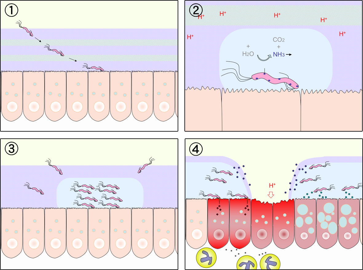
Helicobacter species usually inhabit the gastric mucosa of mammals.
Helicobacter pylori has been shown to be associated with a spectrum of gastrointestinal diseases, including chronic gastritis, gastric and duodenal ulcers, gastric adenocarcinoma and non-Hodgkin's lymphoma of the stomach.
This bacterium colonizes the stomach of over half the world's population, but causes obvious clinical disease in only 5-10% of all infected individuals.
H. pylori is pathogenic because it colonizes the stomach, usually for years or decades, by invading the gastric mucosa and causing a continual low grade inflammation.
DISTINCTIVE PROPERTIES:
Helicobacter pylori is a Gram-negative, helically shaped bacterium that lives in the mucus layer above the gastric epithelium where it is buffered from the highly acidic environment of the stomach.
H. pylori possess multiple flagella at one pole (lophotrichous) and are motile.
The organisms are nutritionally fastidious and microaerophilic, growing ideally in an atmosphere of about 5% oxygen.
H. pylori is a strong producer of urease, an enzyme that catalyzes the hydrolysis of urea into ammonia and carbon dioxide. The organism is also oxidase and catalase positive with optimal growth pH of 6.0-7.0.
The endotoxin of H. pylori is less potent than that of most gram-negative bacterium.
PATHOGENESIS:
Helicobacter pylori ulcer diagram GNU Free Documentation License
The specific mode of transmission and mechanism of H. pylori is unknown, however, direct person-to-person transmission is suspected.
H. pylori may be spread either by the fecal-oral or oral-oral route. The organism has been cultured from the stools of infected children, and from dental plaque, and its DNA has been detected in saliva using Polymerase Chain Reaction.
Essentially, all persons infected with H. pylori develop chronic superficial gastritis involving the antrum and the fundus of the stomach. Infected persons may develop peptic ulcer disease, lymphoproliferative disease, chronic atrophic gastritis and gastric adenocarcinoma years to decades after initial infection.
H. pylori infection is associated with chronic gastric inflammation. Inflammatory effector molecules released by the organisms induce tissue inflammation and release of nutrients. This process of inflammation may serve as a means for the organism to obtain constant nutrients.
H. pylori infection results in the down regulation of somatosatin-producing D cells, which in turn leads to elevated gastrin levels and enhanced gastrin acid secretion and eventually ulceration at the site.
A carrier of H. pylori usually has no symptoms, but persons with gastritis or ulcers may have the following symptoms: abdominal pain, dyspepsia or indigestion, bloating and fullness, mild nausea, belching and regurgitation, weight loss and hunger every 1-3 hours.
Research has indicated that H. pylori infection may lead to gastric cancer via a series of events: chronic superficial gastritis → atrophic gastritis → atrophy → incomplete intestinal metaplasia → dysplasia and finally adenocarcinoma.
HOST DEFENSES:
Gastric acid plays an important role in protection against many other enteric pathogens but is not very effective in preventing colonization of the mucosa by H. pylori.
With the onset of acute infection, a peak in IgM antibodies are first observed followed by peaks in titers of IgA and IgG antibodies. This inflammatory response is ultimately reduced to a low level stable state and tends to persist for years or even decades with most infected persons remaining asymptomatic for life.
While both humoral and cellular recognition of the bacterium occurs, it evades immunological clearance.
EPIDEMIOLOGY:
Persons having frequent contact with human feces and secretions, such as adults living in institutions and children living in orphanages, have the highest infection rates.
Age also seems to be a factor in H. pylori infection; less than 20% of persons under the age of 30 display infection, while persons 60 years and older display a 40-60% infection rate.
Developing countries tend to have a higher prevalence of H. pylori than developed countries - the infection is usually acquired in early childhood.
DIAGNOSIS:
Clinical: Clinical diagnosis of H. pylori infection is often difficult because the symptoms closely resemble that of ulcers, gastric cancers, acid reflux disease, etc. Isolation of the organism is required for positive identification.
Laboratory:H. pylori can be visualized microscopically or cultured on chocolate agar in a microaerophilic environment. After about a week, small, translucent colonies can be tested for urease, oxidase and catalase positive reactions. An in vivo test for urease involves ingestion of 13C or 14C-labeled urea by the patient. If the enzyme is present, the labeled urea with be hydrolyzed to CO2 and 2 NH3; the carbon label will be within the CO2 and this can be detected in the patient's breath.
CONTROL:
Sanitary: Since H. pylori is thought to be acquired via the fecal-oral and the oral-oral route, sanitary means of control are most important.
Immunological: Various vaccines for the treatment of H. pylori infections are currently being developed; some of these vaccines target the motility of the organism, while others the protective membranes.
Chemotherapeutic: The most commonly recommended therapy for H. pylori infection has been triple therapy consisting of a proton pump inhibitor (PPI), amoxicillin, and clarithromycin.